

Product |
Growth Factor |
Phenol Red |
Recommended Applications |
Specifications |
Cat.NO |
GelNest™ Matrix |
Normal |
Yes |
General 2D, 3D cell culture |
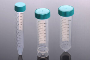
5mL/bottle, 1 bottle/box |
211212 |
GelNest™ Matrix, without Phenol Red |
Normal |
No |
Requires colorimetric identification (such as fluorescence) or sensitivity to steroids |
5mL/bottle, 1 bottle/box |
211222 |
GelNest™ Matrix, low growth factor |
Low |
Yes |
2D, 3D cell culture with higher accuracy requirements for matrix components and requires |
5mL/bottle, 1 bottle/box |
211232 |
GelNest™ Matrix, low growth factor, without Phenol Red |
Low |
No |
colorimetric identification or sensitivity to steroids |
5mL/bottle, 1 bottle/box |
211242 |
GelNest™ Matrix, high concentration |
Normal |
Yes |
In vivo tumor formation, thrombosis test, angiogenesis experiment, general cell culture, etc. |
5mL/bottle, 1 bottle/box |
211252 |
GelNest™ Matrix, high concentration, without Phenol Red |
Normal |
No |
2D, 3D cell culture with higher accuracy requirements for matrix components |
5mL/bottle, 1 bottle/box |
211262 |
GelNest™ Matrix, for stem cells |
Low |
Yes |
hESC stem cell culture |
5mL/bottle, 1 bottle/box |
211272 |
GelNest™ Matrix, for organ-like structures, without Phenol Red |
Low |
No |
Organoid culture and differentiation |
5mL/bottle, 1 bottle/box |
211282 |

NEST® GelNest™ Matrix
Prijsklasse: € 171,25 tot € 288,95 Exclusief BTW
- Accessoires
- Product omschrijving
- Features
- Technical specifications
- Beschrijving
- Extra informatie
- Beoordelingen (0)
GelNest™ Matrix
GelNest™ Matrix is derived from mouse tumor tissue and contains extracellular matrix components such as laminin, type IV collagen, heparan sulfate proteoglycans, and more. These components support cell adhesion, differentiation, and proliferation, providing signals for these processes. Additionally, they simulate the characteristics of the basement membrane in the physiological environment, enhancing the success rate and effectiveness of cell culture.
In addition to the matrix components, GelNest™ Matrix is rich in various growth factors. These growth factors promote cell differentiation, proliferation, and migration, mimicking cellular signaling pathways and interactions in the physiological environment. GelNest™ Matrix has a wide range of applications, particularly in tissue engineering, cell culture, and research. It can be used for organoid culture, stem cell differetiation, angiogenesis, migration or invasion assays, and in vivo tumor studies.
Introduction
Organoid Culture Test
-
Re-suspend the single-cell suspension for organoid culture in pre-cooled basal medium at 4°C and perform cell counting.
-
Mix the cells with the gel solution and add the mixture to pre-warmed 24-well plates. Each well should contain approximately 5×10⁴ cells and 60µL of matrix gel.
-
Immediately place the plates in the incubator, and the gel will solidify in approximately 10 minutes.
-
Add 500µL of organoid culture medium for cultivation.
-
Wait for 3-5 days for the organoids to form. Finally, image the live cells using high-content microscopy to determine the sensitivity of the organoids to various drugs.
This method provides an efficient and convenient solution for organoid culture and can be used in areas such as drug screening and tumor research.
Stem Cell Differentiation Test
For human embryonic stem cells (hESCs) and induced pluripotent stem cells (iPSCs) without feeder layer culture:
-
Thaw GelNest™ Matrix stored in freezing conditions and let it thaw overnight in a 4°C ice bath. Gently blow and mix the gel three times with a pre-cooled pipette tip for thorough mixing. Transfer the thawed matrix gel using the pre-cooled pipette tip. If bubbles form, briefly centrifuge with a handheld centrifuge to remove them.
-
Preheat the cell culture plates in the incubator.
-
Dilute the gel solution in pre-cooled serum-free medium at a 1:100 ratio. Make sure to cover the entire culture plate with the diluted gel solution. The recommended amount is 300µL/cm^2 in the culture dish.
-
Leave the culture plates with the modification solution at room temperature for 1 hour.
-
Remove the modification solution and immediately seed the stem cells mixed with mTeSR in the culture plate. Be cautious to prevent the surface of the modified culture plate from drying out.
This method provides an efficient and convenient solution for stem cell culture and is expected to play an important role in tissue engineering, regenerative medicine, and other fields.
In Vitro Blood Vessel Formation Test
-
Replace the complete growth medium with starvation medium for cells: DMEM medium containing 0.2% FBS, 2mM L-glutamine, 1mM sodium pyruvate, 100U/mL penicillin, and 100µg/mL streptomycin. Starve the cells for 24 hours.
-
Spread 50µL of GelNest™ Matrix evenly on the bottom of a 96-well plate. To prevent the gel from adhering to the pipette tip, aspirate and blow FBS once in the pipette tip to wash the inner wall of the pipette tip with FBS.
-
Place the 96-well plate in a 37°C cell culture incubator and incubate for 30 minutes to solidify the gel.
-
Digest the endothelial cells and perform cell counting.
-
Add 5×10^4 HUVEC cells to the 96-well plate containing the gel, totaling 200µL of cell suspension. Place the 96-well plate in the incubator for cultivation.
-
Vascular-like network structures will form within 3 to 12 hours. This is the optimal observation time.
-
At the optimal observation time, carefully remove the culture medium and stain with culture medium containing a 1/1000 concentration of Calcein AM (green). Image the cells using a microscope and record the morphology and characteristics of the vascular network.
This experiment can be used in the fields of angiogenesis and cardiovascular disease research.
Cell Invasion Test in Cell Culture Insert
-
HT-1080 cells cultured in MEM medium supplemented with 10% fetal bovine serum (FBS) are used in this experiment. When the cell density reaches 80% to 90%, take 20µL of GelNest™ Matrix Gel and dilute it 50 times with serum-free MEM to a final volume of 1000 µL. Gently blow and mix the matrix gel with a pipette for thorough mixing. Add 100µL of the diluted gel mixture to the center of the insert, evenly covering the surface of the insert with the gel mixture. Incubate the culture dish at 37°C for 1 hour to allow gel formation.
-
After trypsinization of the cells (generally, for a 6-well plate, digest with 200µL of trypsin at 37°C for 3 minutes, then terminate the digestion with 10% serum and centrifuge at 300g for 3 minutes), resuspend the cells in serum-free MEM and perform cell counting. Take 750µL of cell suspension at a starting concentration of 1 x 10⁶/mL (estimated to be 10 wells, with 7.5×10⁴ cells per well, totaling 750,000 cells), and dilute with serum-free MEM to a final volume of 1.5mL. Then, add 150µL of cell suspension to the upper chamber of each insert, resulting in 7.5×10⁴cells/well. For the experimental group, 800µL of medium containing 10% fetal bovine serum (FBS) as a chemoattractant will be added to the lower chamber. In contrast, the control group will receive 800µL of medium without FBS in the lower chamber. Culture the cells in a humidified incubator at 37°C and 5% CO2overnight.
-
Discard the culture medium in the inserts and wash twice with PBS. Then, stain the cells on the lower surface of the membrane with crystal violet for 10 minutes. Subsequently, wash the inserts twice with PBS to remove unbound crystal violet. Gently remove the cells inside the inserts with a wet cotton swab and allow them to air-dry. Observe and image the invaded cells under a microscope.
To elute the bound crystal violet, dilute acetic acid to 33% (v/v) with ddH2O. Add 400µL of 33% acetic acid to each insert and shake on a shaker for 10 minutes. Next, transfer the eluent from the lower chamber to a 96-well transparent microplate. Measure the absorbance at 590nm using an ELISA reader.
Figure 4. Results of HT-1080 cells cultured on competitor’s matrix gel-modified surface and GelNest™ Matrix Gel-modified surfaces forming a vascular network after 9 hours. Scale bar: 200µm. The results indicate that FBS can significantly induce cells to penetrate the semi-permeable membrane of the extracellular matrix and enter the lower surface of the insert.
GelNest™ Matrix Quality Assurance
Protein Concentration
The protein concentration is guaranteed to be between 8~20mg/mL.
Stable Performance
The gel formation performance remains stable.
High Safety Performance
There are no LDEV (Lactate Dehydrogenase Elevated Virus), bacteria, or mycoplasma present.
Endotoxin Level
The endotoxin level is <10EU/mL.
Cell Culture
Tests for organ-like structures and stem cell culture have been successfully performed.
Experimental Testing
Tests for angiogenesis, tumor invasion, and tumor formation have been successfully conducted.
GelNest™ Matrix Selection Guide
Download your brochure here: GelNest
Beoordelingen
Er zijn nog geen beoordelingen.